Download The Temporal Bone: Anatomical Dissection and Surgical Approaches - Mario Sanna | ePub
Related searches:
Temporal bone: Anatomy, parts, sutures and foramina Kenhub
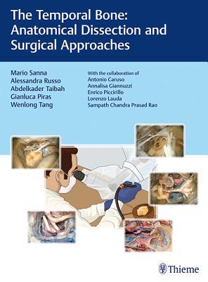
The Temporal Bone: Anatomical Dissection and Surgical Approaches
Temporal bone: Anatomical diagram, function, and injuries
Temporal Bone: Anatomy, Function, and Treatment
(PDF) The Temporal Bone: Anatomical Dissection And Surgical
Temporal Bone Anatomy - Mass. Eye and Ear Otopathology Laboratory
Temporal Bone Anatomy (Cadaveric Dissection) Iowa Head and
The Temporal Bone: Anatomy and Common Pathologies - netkey.at
Temporal Bone Vascular Anatomy, Anomalies, and Disease, with
The Temporal Bone: Anatomical Dissection and - Gruppo Otologico
Temporal Bone Anatomy and Function The House Institute
Temporal Bone Ct and Mri Anatomy - Did You Check eBay
Anatomy of temporal bone and it’s surgical importance
Topographical anatomy and morphometry of the temporal bone of
The Temporal Bone A Manual For Dissection And Surgical - NACFE
1 Anatomy and Radiology of the Normal Temporal Bone Ento Key
Temporal Bone and Ear Anatomy - Kevin Kavanagh MD
Surgical Anatomy of the Temporal Bone and Measurements of the
Home - Mass. Eye and Ear Otopathology Laboratory
Temporal Bone: Definition, Anatomy, and Fracture Micro B Life
Anatomy of the Temporal Bone, External Ear, and Middle Ear
Temporal Bone Ct and Mri Anatomy on eBay - ebay.com
The temporal bone: parts, side dtermination and it's importance
The Skull Anatomy and Physiology I
Skull Anatomy - Cranial Bone and Suture Labeled Diagram
The temporal bone consists of the lateral skull base, forming portions of the middle and posterior fossa (a hollow space in the skull, near the brainstem and cerebellum). The five osseous components of the temporal bone are the squamous, mastoid, petrous, tympanic, and styloid portions(6).
Temporal bone normal anatomy computed tomography /histology correlation otopathology laboratory department of radiology massachusetts eye and ear otopathologylaboratory.
And tympanic parts of the temporal bone have enjoyed increasing popularity in the last two decades1,6,7,11,12,14,15,19-23). Key to understanding these transtemporal approaches and to the in-novation of any new approaches is an under-standing of the anatomy of the temporal bone (fig.
Temporal bone dissection using a human cadaver is the gold standard educational program; however, human cadavers are limited and not available for all medical staff members who want to understand the temporal bone anatomy�.
The temporal bone consists of four named portions: temporal squamosa; tympanic segment (ring housing ear canal) mastoid; petrous portion (pyramid) from the lateral aspect the largest portion seen is the temporal squamosa (squamous part) which occupies the superior two thirds of the illustration.
Temporal bone anatomy is arguably the most complex anatomy in the human body. The proximity of vital neural and vascular structures, the intricate three-dimensional relationships involved, and the manner in which these structures are encased in a labyrinth of bony canals pose a major challenge to the preparation for and performance of surgery.
Temporal bone is an even bone that forms the sides of the skull or temples. It is an irregular bone and lies inferiorly to the cranial casing.
The temporal bones are situated at the sides and base of the skull, and lateral to the temporal lobes of the cerebral cortex. The temporal bones are overlaid by the sides of the head known as the temples, and house the structures of the ears.
7 nov 2019 the temporal bone is one of the thickest bones in the skull. In this article, we look at the structure and function of this bone and the injuries that.
The temporal line of the frontal bone is the continuation of the line formed by the union of the superior and inferior temporal lines of the parietal bone. A temporal surface of a bone is a part of a bone, which contributes to the formation of the temporal fossa.
The preoccipital notch is an indentation in the inferior temporal gyrus, about 3 cm anterior to the occipital pole, formed by the petrous part of the temporal bone. A straight line drawn from the parietooccipital sulcus to the preoccipital notch defines the anterior border of the occipital lobe on the lateral aspect of the hemisphere.
The mastoid part of the temporal bone is its posterior component. The inferior projection of the mastoid part is called the mastoid process.
It is located behind the ear, and is known as the c1 bone of the spinal vertebral level. The mastoid process bone itself is in the shape of a pyramid that projects behind the temporal bone. The temporal bone is located at either side of the skull beneath the temple.
Initial study of the pig`s temporal bone anatomy in order to enable a new experimental model in ear surgery.
The temporal bone is a dense complex bone that constitutes the lower lateral aspect of the skull and has complex anatomy because of the three-dimensional.
Temporal bone anatomy is complicated, as the structure of this paired bone is very irregular.
The temporal bone is one of the most important calvarial and skull base bones.
The temporal bones are a pair of bilateral, symmetrical bones that constitute a large portion of the lateral wall and base of the skull� they are highly irregular.
The temporal bones are paired bones that help make up the sides and base of the skull (cranium). This places them lateral—to the side of—the temporal lobes of the brain’s cerebral cortex, ensuring that the cranium is properly supported and protecting the important structures there.
Understanding the anatomy of the temporal bone is critical to a number of open skull base approaches. A number of critical neurovascular structures, namely, the lower seven cranial nerves and the major vessels to and from the brain, traverse the temporal bone.
Find information on temporal bone removal and processing as well as access to the national temporal bone and the mouse cochlea gene databases. Educational resources discover helpful resources from downloadable 3-d models and simulators to webcasts and publications.
Anatomysurgery of the ear and temporal bonemanual of practical anatomy: previous publications have addressed the topic of temporal bone anatomy, none.
Anatomy in both the axial and coronal first of six axial bone ct images of the left temporal bone presented from superior to inferior shows the labyrinthine.
The anatomical complexity of the temporal bone represent s a challenge in the interpretation of anatomical details and the diagnosis of different pathological condition s in this region.
Temporal bone the middle ear consists of the tympanic cavity and the antrum. The antrum is a large aircell superior and posterior to the tympanic cavity and connected to the tympanic cavity via the aditus ad antrum.
This 3 part 3d printed model derived from ct data highlights the complex anatomy of the temporal bone including bone ossicles, canals, chambers, foramina.
Hrct of temporal bone predicts certain normal anatomical variants of surgical significance preoperatively.
The temporal bone: anatomical dissection and surgical approaches ebook: sanna, mario, russo, alessandra, taibah, abdelkader: amazon.
The temporal bones are a pair of bilateral, symmetrical bones that constitute a large portion of the lateral wall and base of the skull. They are highly irregular bones with extensive muscular attachments and articulations with surrounding bones.
Publicationdate january 15, 2016 this is an updated version of the 2007 article. In this review we present the normal axial and coronal anatomy of the temporal bone by scrolling through the images.
The temporal bone has a pyramidal shape, the sides of which form the middle fossa floor (superior face), the anterior limit of the posterior fossa (posterior face), muscle attachments of neck and infratemporal fossa (anteroinferior face), and the muscular-cutaneous–covered side of the head (lateral), which forms the base of the pyramid.
To create the zygomatic arch it joins the temporal process of the zygomatic bone. A deep cavity behind the articular eminence is known as mandibular fossa. The mandibular fossa and articular tubercle are essential portions of the squamous part of the temporal bone. Internal surface is located in contact together with the temporal lobe of the brain.
The temporal bones are two major bones in the skull, or cranium. They help form the sides and base of the skull, where they protect the temporal lobe of the brain and surround the ear canal.
The ossicles located within the middle ear cavity are the three smallest bones in the human body. The transmit and amplify sound from the tympanic membrane to the oval window. The malleus is the most lateral in position and connects to the tympanic membrane.
Temporal bone anatomy is complex, and further complicated by the small size and three-dimensional orientation of associated structures. Computed tomography (ct) has revolutionized imaging of the temporal bone.
The temporal bone or os temporale is a paired, irregular bone and the thickest in the human body, located at the sides and base of the skull. It provides space for important cranial arteries, veins, and nerves. The os temporale also provides attachment points for numerous muscles.
Since 1967, anson and donaldson's surgical anatomy of the temporal bone has been the gold standard reference in its field. Duckert, lambert, and rubel--all preeminent leaders in otolaryngology-head and neck surgery--dr. Donaldson has further refined this classic text/atlas to make it even more useful for study, reference, and presurgical review.
The temporal bone has a pyramidal shape, the sides of which form the middle fossa floor (superior face), the anterior limit of the posterior fossa (posterior face), muscle attachments of neck and infratemporal fossa (anterior-inferior face) and the muscular-cutaneous-covered side of the head (lateral) which forms the base of the pyramid.
The temporal bone consists of four parts— the squamous, mastoid, petrous and tympanic parts. The squamous part is the largest and most superiorly positioned.
Anatomical structure the temporal bone itself is comprised of five constituent parts. The squamous, tympanic and petromastoid parts make up the majority of the bone, with the zygomatic and styloid processes projecting outwards.
For this purpose, dry skulls with intact ossicles were scanned in axial and coronal projections. The detailed ct anatomy of the temporal bone was documented,.
Methods of preparation and study of temporal bones; techniques of removal of human temporal bones; national temporal bone database; educational resources. Downloadable 3-d models; simulator for temporal bone surgery; otopathology webcasts; research fellowships; publications; temporal bone anatomy; useful external links; image libraries.
This paper describes the anatomy of the chinchilla's temporal bone, and four surgical approaches to the labyrinth and ossicular chain, three through the bulla.
15 jan 2016 in this review we present the normal axial and coronal anatomy of the temporal bone by scrolling through the images.
Conclusion: hrct temporal bone delineates the location and extent of the disease and provides information on anatomical variations and complications. Keywords: high-resolution computed tomography temporal bone, radiology in cholesteatoma, unsafe chronic suppurative otitis media.
The anatomyguy site uses cadaver materials and surgical footage which may be considered disturbing to some viewers.
The temporal bone is the bony framework within which a complex array of anatomic relationships exists. These anatomic relationships involve important vascular structures, including the sigmoid sinus, internal jugular bulb/vein, and petrous segment of the internal carotid artery (ica) and the closely associated nervous structures.
The temporal bones are situated at the sides and base of the skull.
Anatomy of the temporal bone with surgical implications, by schuknecht and gulya, is a welcome addition to our educational resources.
Detailed knowledge of the complex microanatomy of the temporal bone is essential for surgeons executing invasive therapeutic procedures.
1 anatomy and radiology of the normal temporal bone basic anatomical knowledge of important structures that may be encountered during middle ear surgery is described here. Since three-dimensional anatomy of the middle ear is so complicated, it is impossible to figure out its entity only through these flat pictures.
The temporal bones of adult balb/c mice were examined and 3d high-resolution reconstructions of the temporal bone were obtained using a micro-ct system. Using the system described here, the bony labyrinth and membranous labyrinth could be investigated in a non-destructive manner.
Based on the dissections of 24 bones of 12 macaques (macaca mulatta), a systematic anatomical description was made and measurements of the cho- sen size.
About the temporal bone of the pig and its use as an experimental model for otological surgery training. Because of this, the aim of this study is to perform an initial study of the pig`s temporal bone anatomy in order to enable a new experimental model in ear surgery.
Everyone has two of these bones, one on either side of the skull, with the bones comprising part of the sides and base of the skull. These bones are closely involved in the anatomy of the ear, and they house a number of anatomical structures of importance.
Publicationdate july 15, 2006 updated version: 21-2-2007 in this review we present the normal coronal and axial anatomy of the temporal bone.
The temporal bones are situated at the sides and base of the skull. The squama, the petrous, mastoid, and tympanic parts, and the styloid process.
Illustration of transversal section of the petrous portion of the temporale bone.
Erik beek temporal bone� the middle ear scroll through the axial anatomy from inferior to superior.
Method: fourteen temporal bones were dissected in the anatomical laboratory of professor edmundo vasconcelos hospital, são paulo-brazil.
Temporal bones the temporal bones are next in the mnemonic, and one of them is shown in orange below. There are 2 temporal bones, one located on either side of the skull in the “temple” region.
The temporal bone is a paired symmetrical bone which forms the lower lateral walls of the human skull. It is a composite structure consisting of four parts, each ossifying independently and later fuses. The temporal, parietal and frontal bones together forms the pterion, which is the weakest part of the skull.
A prominent feature of the temporal bone is this large projection, the mastoid process. As we'll see, it's the origin of some of the muscles that move the head,.
11 may 2018 the primary purpose of temporal bone drilling is to learn temporal bone anatomy. Figure 1 shows a near-complete cortical mastoidectomy (with.
Interactive atlas: this atlas allows you to scroll through ct slices of the temporal bone in four different planes.
Surgical instruments; general guidelines for drilling; suction irrigation; preparation of the specimen; temporal bone holder; anatomy of the temporal bone. Squamous bone; tympanic bone; mastoid process; petrous bone; the middle ear; internal auditory canal; the intratemporal facial nerve; endoscopic surgical.
The anatomy of the temporal bone is one of the most complicated areas in the human body.
The temporal bone (latin: os temporale) is a paired bone situated at the lateral side and base of the skull.
Diseased and normal temporal bones within the same patient, our study primarily recorded the prevalence of five normal variants of the temporal bone. In this regard, there was no difference in the prevalence of anatomical variations studied between the right or left temporal bones.
28 jun 2017 introduction to temporal bone anatomy it is anatomically divided into four regions called the squamous, mastoid, temporal, and petrous parts.
Hrct temporal bone despite its pitfalls such as more radiation exposure and higher cost delineates the location and extent of the disease and provides information on anatomical variations and complications. It really serves as road map to assist the surgeon during surgery.
Temporal bone and ear anatomy: the flash presentation and web pages contained in this section present 60 temporal bone anatomy slides. Histology sections from both the right and left temporal bone are available.
Objectives: this study aimed to evaluate the prevalence of normal variations of temporal bone anatomy on high-resolution computed tomography imaging and report their clinical importance. Methods: a retrospective review was conducted of high-resolution temporal bone computed tomography imaging.
The left temporal bone has better orientation with the gross anatomy better demonstrated. View left temporal bone slides the basilar membrane, utricle and saccule are seen in most specimens. In the performance of a stapedectomy, the stapes footplate is fixed and must be removed.
There are grooves in this surface called sulci (a depression that something runs in): ex: temporal arteries run in sulci which is extra protection from the bone. Sulcus for middle temporal artery the sulcus for the middle temporal artery passes upward on the posterior part of the squama.
Post Your Comments: